
 French
 English
 Chinese
 Russian
MSC Therapy for Multiple Sclerosis: 2026 RCT Data & Meta-Analysis
Multiple sclerosis (MS) affects approximately 2.8 million people worldwide, with progressive forms—primary progressive MS (PPMS) and secondary progressive MS (SPMS)—representing the hardest clinical challenge. Unlike relapsing-remitting MS, where dozens of disease-modifying therapies exist, treatment options for progressive MS remain limited. Ocrelizumab offers modest benefit in PPMS, and siponimod is approved for active SPMS, but for patients with non-active progressive disease, no approved therapy slows disability accumulation. Mesenchymal stem cells (MSCs) offer a dual mechanism worth investigating: immunomodulation to suppress the autoimmune attack on myelin, and neurotrophic support to protect vulnerable neurons. Three 2026 publications—the SMART-MS RCT, a meta-analysis of RCTs, and the MESEMS Phase 2 immunological substudy—provide the most rigorous clinical data to date on MSC transplantation in progressive MS.
The SMART-MS Trial: First Rigorous Placebo-Controlled RCT
Published in Neurology by Kvistad et al. (2026), SMART-MS (NCT04749667) is the highest-quality evidence available on MSC therapy for progressive MS. This randomized, double-blind, placebo-controlled Phase I/II crossover trial enrolled 18 patients (mean age 46.7 years, 55.6% female) across four Norwegian hospitals. Participants received a single intrathecal injection of autologous bone marrow-derived MSCs (1 × 10⁶ cells/kg). The primary endpoint was combined evoked potentials—a sensitive measure of CNS conduction that detects subclinical changes even when disability scores remain stable.
Key Findings
- Primary Endpoint: No significant between-group difference in evoked potential latency at 6 months (β = -0.31, 95% CI -1.84 to 1.22, p = 0.668). This Class III evidence indicates a single intrathecal MSC dose does not produce a measurable neuroregenerative effect on neural conduction.
- MRI Cerebral Atrophy: The MSC group showed reduced cerebral atrophy at 6 months (β = 9.37, 95% CI 0.29 to 18.45, p = 0.044). Brain atrophy is a hallmark of progressive MS and correlates with long-term disability. However, this effect was not sustained at 12 months, suggesting a transient protective effect from a single dose.
- Serum Biomarkers: Serum glial fibrillary acidic protein (GFAP), a marker of astrocytic damage, showed a trend toward reduction at 6 months (β = -16.3 pg/mL, 95% CI -33.0 to 0.3, p = 0.054).
- CSF Proteomics: Exploratory CSF analysis revealed reductions in multiple inflammation-related proteins at 6 months, including innate immune activation markers and complement pathway components.
- Safety Signal: Nine patients developed post-procedural fever, ten reported low back pain, and seven showed spinal MRI abnormalities (fluid loculations, nerve root clumping) at 6 months. One patient developed chronic coccygeal pain attributed to arachnoiditis. One serious adverse event was deemed probably related to MSC treatment.
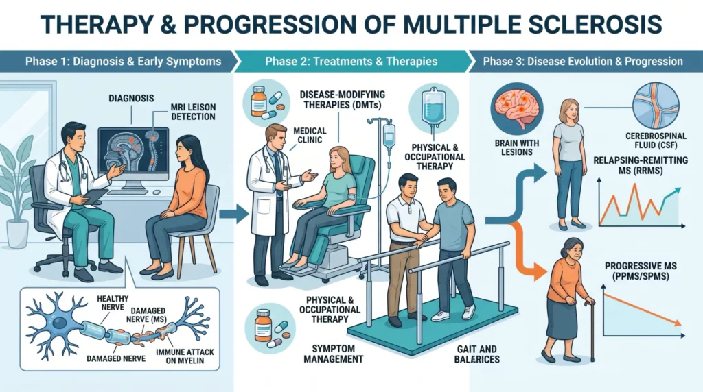
Meta-Analysis of RCTs: Pooling the Evidence
Guo et al. (Frontiers in Immunology, 2026) conducted a systematic review and meta-analysis pooling exclusively RCTs of MSC transplantation in PMS. The search covered multiple databases through March 2026, following PRISMA 2020 guidelines (PROSPERO: CRD420261301154).
Three RCTs met inclusion criteria: 143 patients (75 MSC, 68 control). Primary outcome was EDSS score change. Key results:
- EDSS Score: No significant improvement (pooled MD = -0.08, 95% CI -0.38 to 0.22, P = 0.61). Heterogeneity was moderate (I² = 66%), reflecting differences in MSC source, dose, and delivery across trials.
- Adverse Events: MSC transplantation did not increase TEAEs (RR = 1.08, P = 0.52) or SAEs (RR = 0.79, P = 0.67).
- Certainty: GRADE rated moderate certainty for TEAE safety, very low certainty for EDSS change and SAEs.
The conclusion: MSC transplantation appears safe but does not improve EDSS scores in unselected PMS patients. The qualifier “unselected” becomes central when interpreting the MESEMS immunological data.
MESEMS Phase 2: Immunological Biomarkers
The MESEMS trial (Transplantation and Cellular Therapy, 2026) by Schiavetti, Freedman, and colleagues was an international randomized, double-blind, placebo-controlled Phase 2 crossover study. While the primary MRI endpoint was not met, this ancillary study provides the most detailed immunophenotyping data available from an MSC trial in MS, analyzing peripheral immune changes in 51 patients.
Immune Cell Dynamics
- CD8+ T Cells: MSC treatment increased CD8+ T cells (P = .032). These cells play a complex role—they can be both pathogenic (cytotoxic attack on oligodendrocytes) and regulatory (CD8+ Tregs suppress autoimmune responses).
- CD19+ B Cells: A relative reduction in CD19+ B cells occurred within 4 weeks (estimate -1.97%, 95% CI -3.43 to -0.50, P = .033). B-cell depletion is a proven MS treatment strategy, and MSC-mediated B-cell suppression may contribute to their effect.
- Regulatory T Cells: CD4+ Tregs increased (P = .006). Tregs are deficient in MS patients, and their restoration is associated with disease stability.
- T-NK-like Cells: In MSC-treated participants, relapses were preceded by decreases in CD3+CD56+ T-NK-like cells (P = .035), whereas placebo relapses were preceded by CD8+ T-cell decreases (P = .021). This differential pattern suggests distinct immune pathways preceding disease activity depending on treatment.
Perhaps the most actionable finding: lower early transitional B-cell delta values predicted subsequent MRI activity in placebo patients (P = .005), suggesting immune monitoring could identify patients at risk of progression—a potential tool for personalized treatment decisions.
Mechanistic Insights
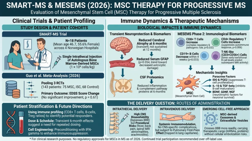
The combined data reveal that MSCs exert their primary effects in MS through immunomodulation rather than direct neuroregeneration. MSCs operate through paracrine signaling rather than direct cell replacement.
- IDO and PGE2 Secretion: MSCs secrete indoleamine 2,3-dioxygenase and prostaglandin E2, which suppress T-cell proliferation and promote regulatory phenotypes by depleting local tryptophan.
- B-Cell Suppression: The CD19+ reduction may be mediated through MSC-secreted IL-10 and TGF-β, which inhibit B-cell maturation.
- Treg Induction: MSCs promote FoxP3+ regulatory T-cell expansion through cell-cell contact and soluble factors, helping restore immune tolerance to myelin antigens.
- Neurotrophic Factors: MSCs produce BDNF, GDNF, and NGF, supporting neuronal survival and oligodendrocyte function. The GFAP reduction in SMART-MS suggests MSCs may dampen reactive astrogliosis.
- Complement Inhibition: CSF proteomics showing reduced complement pathway components suggest attenuation of complement-mediated tissue damage, an increasingly recognized demyelination mechanism in progressive MS.
The Delivery Question
SMART-MS highlights a critical practical issue: route of administration. Intrathecal delivery bypasses the blood-brain barrier for high CNS bioavailability, but carries procedural risks including arachnoiditis. Intravenous delivery avoids CNS-specific complications but introduces the pulmonary first-pass effect, where most MSCs become trapped in lung capillaries. Yet IV delivery provides systemic immunomodulation through circulating paracrine factors—many successful MSC trials in GvHD and Crohn’s fistula have used IV delivery with acceptable safety.
Emerging cell-free approaches—particularly MSC-derived exosomes and extracellular vesicles—may resolve this dilemma. These nano-sized particles cross the BBB more readily than whole cells, carry therapeutic cargo (mRNAs, microRNAs, proteins) without risks of cellular embolization, and can be manufactured with standardized potency assays.
Clinical Implications and Future Directions
The 2026 evidence shows MSCs have clear biological activity in MS—reducing brain atrophy transiently, modulating immune cell populations, and suppressing inflammation-related CSF proteins. Safety is established for IV delivery, though intrathecal administration carries risks the SMART-MS investigators recommend approaching with caution. What the evidence does not yet show is a convincing effect on clinical disability (EDSS) in unselected populations. This gap between biological activity and clinical efficacy points to priorities for the next generation of trials:
- Patient Stratification: The MESEMS biomarker data suggest immune profiling could identify responders. CD8+ T-cell dynamics, transitional B-cell levels, and Treg/effector ratios may serve as predictive biomarkers.
- Dose and Schedule: The transient effects in SMART-MS (present at 6 months, gone at 12 months) argue for repeated dosing. No trial has tested multi-dose regimens in progressive MS.
- Combination Approaches: Pairing MSCs with established DMTs (e.g., ocrelizumab for B-cell depletion synergy) or remyelination-promoting agents could address multiple disease mechanisms.
- Cell Engineering: Preconditioning MSCs with interferon-gamma enhances their immunosuppressive capacity by upregulating IDO and HLA-G expression.
For clinicians considering MSC therapy for progressive MS patients today, the evidence supports continued participation in clinical trials rather than off-label use. The biological rationale is strong, the safety profile for IV delivery is acceptable, but clinical efficacy data remain insufficient to recommend MSCs outside research settings.
Frequently Asked Questions
Is MSC therapy approved for multiple sclerosis?
No. As of 2026, no regulatory agency has approved MSC therapy for MS. All clinical trials remain in Phase I or II. MSCs for MS are available only through clinical trials.
How do MSCs compare to ocrelizumab?
They cannot be directly compared yet. Ocrelizumab has large Phase III data showing reduced disability progression in PPMS. MSC trials are much smaller and have not demonstrated EDSS improvement. MSCs may ultimately serve as adjunctive therapy rather than a replacement for established DMTs.
What are the risks?
For IV delivery, risks appear low—comparable to placebo in the pooled meta-analysis. For intrathecal delivery, SMART-MS documented fever, back pain, spinal MRI abnormalities, and one case of chronic arachnoiditis.
What types of MSCs are used?
Most trials use autologous bone marrow MSCs. The MESEMS and SMART-MS trials both used autologous BM-MSCs. Some trials explore umbilical cord-derived or adipose-derived MSCs, which offer allogeneic off-the-shelf potential with different immunomodulatory profiles.
When will we know if MSCs work for progressive MS?
Larger Phase II/III trials with repeated dosing and biomarker-guided patient selection are needed. Meaningful efficacy data from well-powered trials may emerge within 3-5 years. Several ClinicalTrials.gov registrations for progressive MS MSC trials are active as of 2026.
Références
- Kvistad CE, Kråkenes T, Holmøy T, et al. Intrathecal Mesenchymal Stem Cells in Progressive Multiple Sclerosis: A Randomized, Double-Blind, Placebo-Controlled Trial (SMART-MS). Neurology. 2026;106(10):e214915. doi:10.1212/WNL.0000000000214915
- Guo H, Zeng L, Sun L. Efficacy and safety of mesenchymal stem cell transplantation for progressive multiple sclerosis: a systematic review and meta-analysis of randomized controlled trials. Front Immunol. 2026;17:1861624. doi:10.3389/fimmu.2026.1861624
- Schiavetti I, Freedman MS, Thebault S, et al. Effect of Mesenchymal Stem Cells on Peripheral Immune Cells in Multiple Sclerosis: Results from an International Phase 2 Clinical Trial. Transplant Cell Ther. 2026. doi:10.1016/j.jtct.2026.08.016
- Nahayati MA, Ghavidel Yazdi S, Hassanisabzevar M. Mesenchymal stem cells in clinical trials for multiple sclerosis: A systematic literature review. Mult Scler Relat Disord. 2025;106694. doi:10.1016/j.msard.2025.106694
- Arab FL, Yousefi F, Naderi-Meshkin H, et al. Safety and feasibility of intravenous fresh adipose-derived MSCs in secondary progressive MS: phase I/IIa results. Stem Cell Res Ther. 2026. doi:10.1186/s13287-026-04933-6
- Maji S, Mishra A, Srinivasan A. Efficacy and safety of hematopoietic and mesenchymal stem cells in multiple sclerosis: a meta-analysis. Neurol Res. 2026. doi:10.1080/01616412.2025.2521712